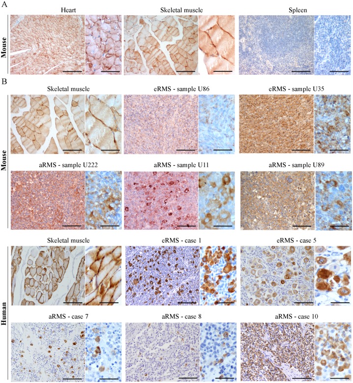
Fig 2. Expression of MURC/cavin-4 in RMS tumors.
A) The specificity of MURC/cavin-4 antibody was tested by IHC analysis using mouse tissue samples derived from heart, skeletal muscle and spleen. The latter was expectedly negative to MURC/cavin-4 staining (brown). Images were taken at 20x and 60x magnification. Scale bars:100 μm. B) MURC/cavin-4 staining (brown) was evaluated by IHC analysis on mouse and human tumors (as reported in Table 1). Skeletal muscle served as a positive control. Representative pictures were taken at 20x and 60x magnification. Scale bars:100 μm.

