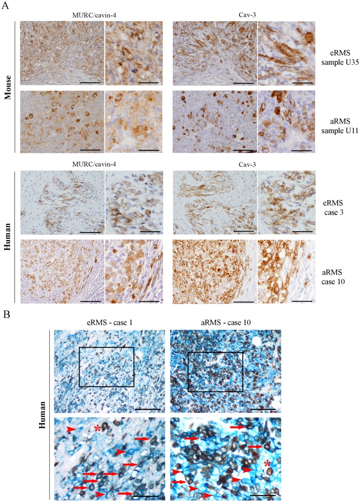
Fig 3. Concurrent expression of MURC/cavin-4 and Cav-3 in RMS tumors.
A) Single staining (brown) of either MURC/cavin-4 or Cav-3 was evaluated by IHC analysis on serial tumor sections established from mouse and human tumor samples (as reported in Table 1). Representative pictures were taken at 20x and 60x magnification. Scale bars:100 μm. B) MURC/cavin-4 (brown) and Cav-3 (blue) staining was evaluated by double IHC analysis on human tumors. Representative pictures were taken at 20x magnification, whereas 60x magnification corresponds to inset; scale bars: 100 μm. * corresponds to MURC/cavin-4 brown staining, arrowhead corresponds to Cav-3 blue staining and arrow corresponds to double staining.

