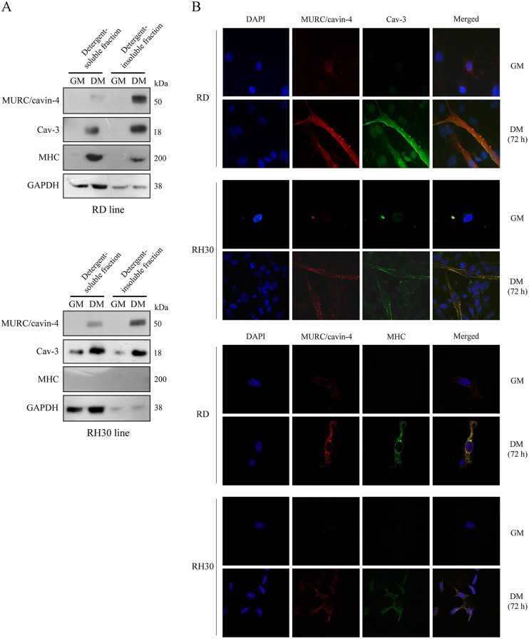
Fig 5. Subcellular localization of MURC/cavin-4 and Cav-3 in the human RD and RH30 lines.
A) Embryonal RD and alveolar RH30 cells were seeded in 60-mm dishes (at a density of 12 x 104) and cultured in GM for 72 hours until reaching confluence; cells were then treated with DM for additional 72 hours. Protein homogenates were subjected to cell fractionation, and the detergent-soluble and-insoluble fractions were analysed by immunoblotting to evaluate the protein levels of MURC/cavin-4, Cav-3, MHC and GAPDH. Results are representative of three independent experiments. B) Confocal microscopy analysis was employed to analyze the distribution of MURC/cavin-4 (red), Cav-3 (green) and MHC (green) in RD and RH30 cells cultured in GM or DM. Nuclei were counterstained with DAPI (blue). Samples were analyzed using a Zeiss LSM510 META microscope equipped with a 63x oil immersion objective. Merged images, captured using the LSM 510 Meta software, showed an extensive co-localization of MURC/cavin-4 with Cav-3 at the cell surface as well as with MHC in the cytosol (yellow signal). Pictures were taken at 63x magnification.

