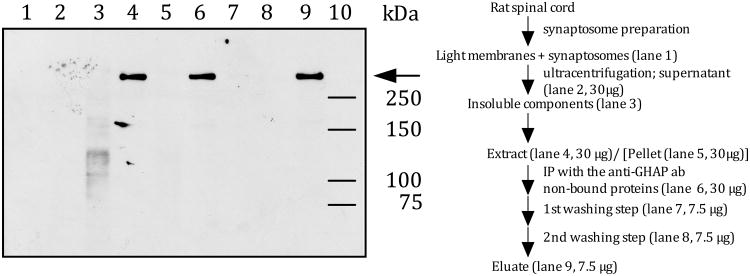
Figure 1. IB4-reactivity in rat spinal cord.
Rat spinal cord tissue was fractionated according to a synaptosome preparation scheme (Gray and Whittaker 1962). A combined light membrane and synaptosome fraction was treated with hyaluronidase to release hyaluronan-bound proteins, such as versican, into the supernatant. Proteins in the hyaluronidase-extract were immunoprecipitated with the monoclonal anti-versican antibody (12C5), which binds all known versican splice variants (Westling et al. 2004).
Different amounts of proteins (see fractionation scheme on the right) from each fraction were separated by SDS-PAGE and electroblotted onto a nitrocellulose membrane. IB4-reactivity (arrow) was visualized using HRP-conjugated IB4 and ECL as the detection system.

