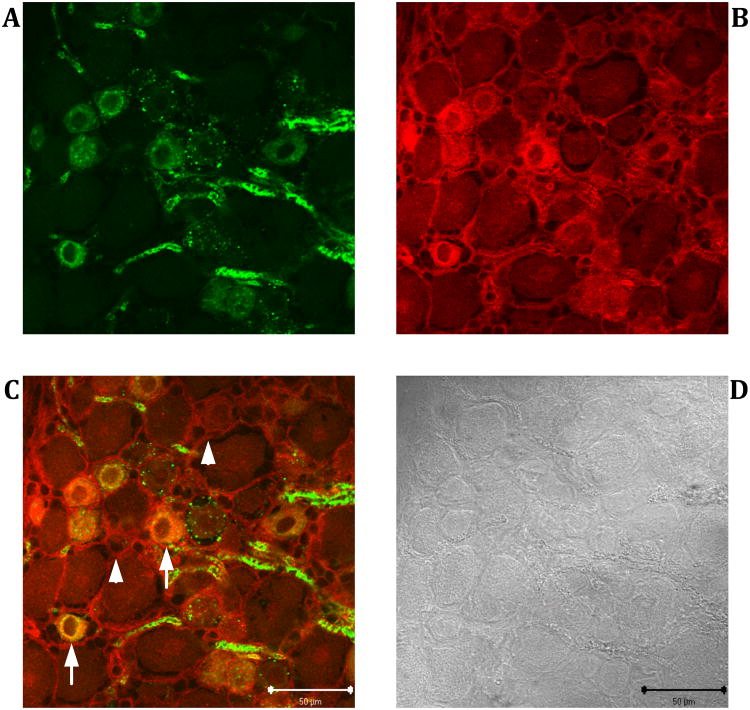
Figure 4. Co-localization of IB4- and anti-versican immunoreactivity in rat L4 DRG.
Dual-labeling immunofluorescence experiments were performed on 20 μm thick cryostat sections. IB4-reactivity (green) was revealed with FITC conjugated IB4 (A), anti-versican immunoreactivity (red) by probing the tissue sections with the mouse monoclonal anti-versican antibody followed by a Cy3-labeled rabbit anti-mouse antibody (B). Subcellular areas in which both immunoreactivities co-localize appear yellow in the merged image (C). Corresponding bright field image captured with differential interference contrast optics (D). Arrows: IB4-binding sensory neurons that also express versican. Arrowheads: Versican immunoreactivity in the extracellular matrix. Scale bar: 50 μm.

