Figure 1.
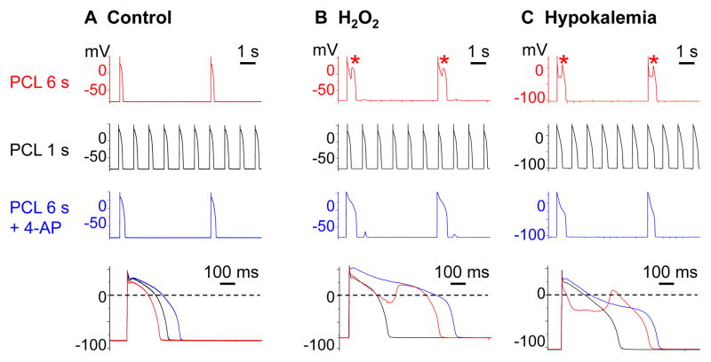
Ito blockade by rapid pacing at PCL 1 s or by 4-AP suppresses H2O2-induced and hypokalemia-induced EADs in rabbit ventricular myocytes. A. No EADs arose under control conditions at PCL 6 s, 1 s, or 6 s in the presence of 4-AP (2 mmol/L). B–C. Following exposure to H2O2 (1 mmol/L; B) or hypokalemia (2.7 mmol/L; C), EADs (*) occurred at PCL 6 s (row 1), but were suppressed by rapid pacing at PCL 1 s (row 2) or by adding 4-AP (row 3). Superimposed action potentials under the 3 conditions are shown below.

