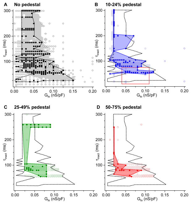
Figure 6.
Virtual Ito parameter combinations causing pacing-suppressed H2O2-induced or hypokalemia-induced EADs to reappear. Graphs show (Ḡto, τinact) combinations that did (solid circles) or did not (open circles) cause EADs to reappear at PCL 1 s, using the protocols shown in Figures 3–5, for the different ranges of Ito pedestal components as indicated in A–D. A. Dashed black line outlines the border of the experimental region in parameter space causing EADs to reappear, compared to the predictions from a computer model (gray shaded area), adapted from Zhao et al.3 B–D. Solid colored regions outline the experimental region in parameter space causing EADs to reappear for pedestals ranging from 10–24% (B), 25–49% (C), and 50–75% (D), compared to the no-pedestal case (black line reproduced from A). In B, the red box indicates the typical parameter values for human ventricular Ito1,f in normal and failing hearts (see Table 1). No EADs re-emerged with pedestals>75%.

