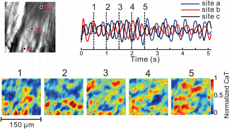
Figure 6.

Random Subcellular Ca2+ oscillations during ventricular arrhythmia. Disorganized subcellular Ca2+ dynamics is occasionally observed under LQT2 conditions, with multiple Ca2+ wavefronts which collide, break and annihilate each other (bottom, 1–5). Ca2+ signal tracings (top right) from 3 pixels indicated in high-resolution image (top left, 150×150 μm2 image size) illustrate the lack of spatial synchrony. Stable Ca2+ baseline is not reached in any cell in the field-of-view, and the signals from different regions of the same cell are poorly correlated. This may reflect an extreme degree of cellular Ca2+ overload. See also Movie G.
