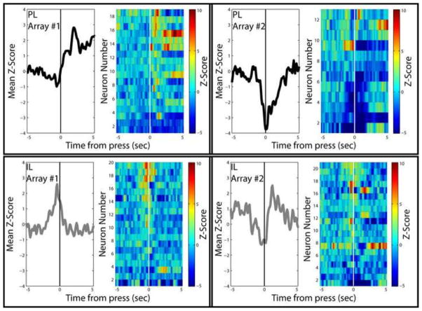
Figure 2.
Example recordings from PL and IL neurons during cocaine self-administration. Each box shows the activity of all neurons recorded from a single array implanted in either PL (top) or IL (bottom) neurons. Each array was implanted in a different rat. Within each box, mean z-scores (left) represent the average of all recorded neurons. Pseudocolor heat plots (right) show the average activity of each neuron. All figures are aligned on cocaine-reinforced lever press. The left two boxes show arrays in PL and IL from which neurons were recorded which were primarily excited at lever press. The right two boxes show arrays in PL and IL from which neurons were recorded which were primarily inhibited at lever press. This figure demonstrates two important details regarding the role of PL and IL neurons in cocaine seeking. First, both PL and IL neurons exhibit both excitatory and inhibitory responses during cocaine self-administration. Second, even within each array, neurons exhibit a variety of responses – some excited, some inhibited, and some nonresponsive – over a wide range of timeframes. These and other data argue against a selective encoding of cocaine seeking in either PL or IL but, instead, indicate that networks within both regions are likely involved (Moorman and Aston-Jones, Submitted-b).

