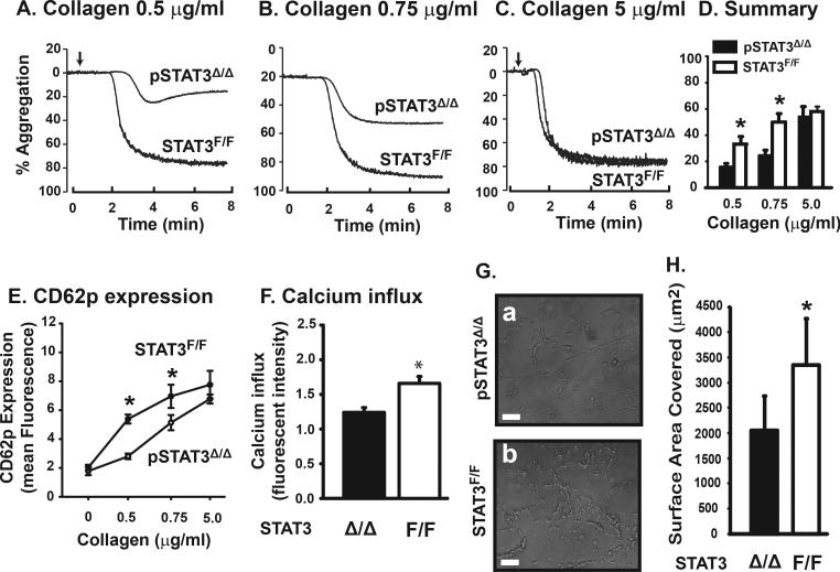
Figure 3.
Platelet function of STAT3Δ/Δ mice: Platelets from pSTAT3Δ/Δ and STAT3F/F littermates were induced to aggregate by 0.5, 0.75, or 5 μg/ml of collagen (A-C) and data from 32 mice/group were quantified (D, *p < 0.01). CD62p expression was measured after platelets were stimulated with 0.5, 0.75 or 5 μg/ml of collagen. The comparison was made between WT and STAT3 KO platelets at each collagen level with a two-way ANOVA (E, n = 12, *p < 0.001). No interaction between litter and genotype was found at any of these collagen doses. Calcium influx was detected in platelets stimulated with 0.75 μg/ml of collagen (F, *p < 0.003). Blood was perfused over immobilized collagen for 10 min at a flow rate of 1 ml/min to measure thrombus formation (G, representative images, bar = 50 μm), which was quantified by measuring surface areas covered by platelet thrombi (H, n = 10, *p < 0.001).

