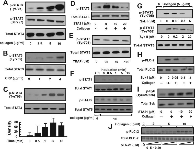
Figure 4.
STAT3 phosphorylation in human platelets: Washed platelets were incubated with various concentrations of collagen (A) or CRP (B) for 10 min at 37°C. Platelet lysates were probed with antibodies against Tyr705 phosphorylated, Ser727 phosphorylated, and total STAT3. Aliquots of platelets were collected and probed for STAT3 phosphorylation over a 15 min after platelets were stimulated with 5 μg/ml of collagen. The optical density of immunoreactive bands of STAT3 phosphorylation was recorded (C). STAT3 phosphorylation induced by 5 μg/ml of collagen was measured in the presence of STA21 (D). STAT3 phosphorylation was also determined in TRAP-treated platelets (E). STAT1 and STAT5 phosphorylation was probed in collagen-stimulated platelet lysates (F). Human washed platelets were first treated with one of two Syk inhibitors for 10 min and then stimulated with 5 μg/ml of collagen. Platelet lysates were probed for the phosphorylation of STAT3 (G) and PLCγ2 (H). STA21-treated platelets were stimulated with collagen and probed for Syk phosphorylation (I). Phosphorylated and total PLCγ2 was probed in platelets treated with various doses of collagen in the presence of increasing doses of STA21 (J). Panel figures represent 3–7 separate experiments.

