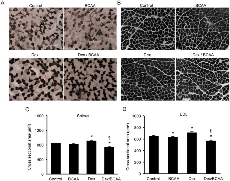
Fig 2. The CSAs of the muscle fibers in the soleus and EDL muscles of the SDRs treated with dexamethasone, BCAA or both.
A. ATPase staining (pH 10.7) of soleus muscle fibers from the SDRs. In this staining, type 1 fibers are stained light, and type 2 fibers are dark. Scale bar: 100 μm. B. ATPase staining (pH 10.7) of EDL muscle fibers from the SDRs. C. BCAA did not increase the CSAs in the soleus muscles. Dex elicited a slight increment in the CSAs compared to the control. The administration of both Dex and BCAA resulted in a decrease in CSA compared to the Dex-treated SDRs. *, P < 0.05 vs. control group; ¶, P < 0.05 vs. Dex-treated group. D. BCAA did not increase but rather decreased the CSAs in the EDL muscles. Dex elicited a slight increment in CSA, and the administration of both Dex and BCAA resulted in a decrease in CSA compared to the control and Dex-treated SDRs, respectively. *, P < 0.05 vs. control group; ¶, P < 0.05 vs. Dex-treated group.

