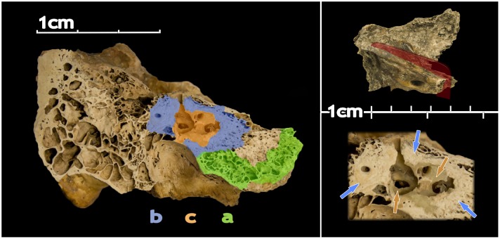
Fig 1. Medial view of a cut of a left petrous bone.
The main image shows the location of the different areas targeted in this study (parts A, B and C) with different colours. The top box shows the direction of the cut. The lower box shows the area comprising parts B and C in detail and non-coloured. Blue and orange arrows point to areas of B and C, respectively.

