
Fig 1. Ultrasonographic grading of pneumonia with necrotizing changes and flow chart of ultrasonograhic assessment.
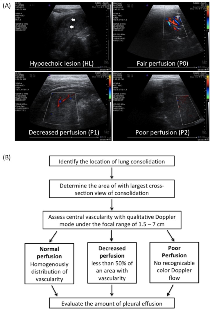
(A) Hypoechoic lesions (HLs), rough-contoured heterogeneously hypoechoic areas in the consolidated lung, are indicated with arrows. Perfusion within the consolidated lung was assessed according to vascularity by using color Doppler. Normal, decreased, and poor perfusion are designated with P0, P1, and P2. (B) Step-by-step flow chart of ultrasonographic assessment.