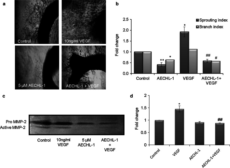
Fig. 4.
AECHL-1 inhibits VEGF-induced microvessel sprouting ex vivo. Aortic segments isolated from Wistar rats were placed in the Matrigel-covered wells and treated with VEGF in the presence or absence of AECHL-1. a Representative images of sprouts from the margins of aortic rings. Images were captured and quantified using Image pro-plus and ImageJ software, respectively. b MMP-2 activity from supernatants assayed using gelatin zymography. Quantification of band intensities was carried out using ImageJ. Columns, mean from three independent experiments; bars, SE. *P < 0.05; **P < 0.01 versus control and # P < 0.05; ## P < 0.01 versus VEGF

