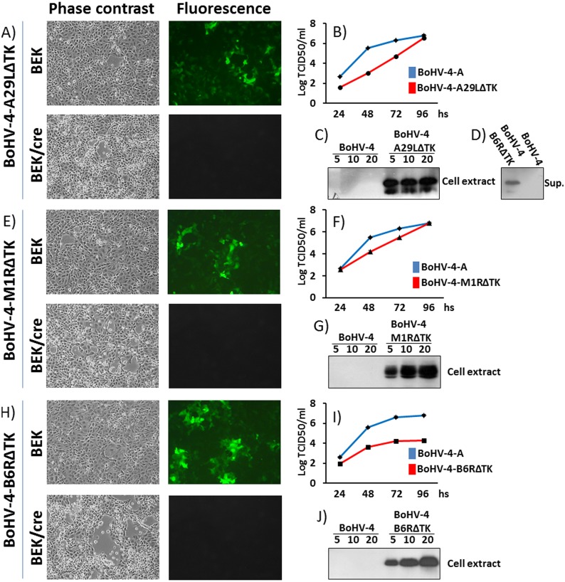
Fig 3. Reconstitution and characterization of recombinant viruses.
Representative phase contrast and fluorescent microscopic images of plaques formed by viable reconstituted recombinant BoHV-4-A-CMV-A29LgD106ΔTK (A), BoHV-4-A-EF1α-M1RgD106ΔTK (E) and BoHV-4-A-EF1α-B6RgD106ΔTK (H) after the corresponding BAC DNA electroporation into BEK cells or in BEK cells expressing cre recombinase (Magnification, ×10). Replication kinetics of BoHV-4-A-CMV-A29LgD106ΔTK (B), BoHV-4-A-EF1α-M1RgD106ΔTK (F) and BoHV-4-A-EF1α-B6RgD106ΔTK (I) growth on BEK cells, compared with those of the parental BoHV-4-A isolate. The data presented are the means ± standard errors of triplicate measurements (P>0.05 for all time points as measured by Student's t test). Western immunoblotting of cells, infected with BoHV-4-A-CMV-A29LgD106ΔTK (C), BoHV-4-A-EF1α-M1RgD106ΔTK (G) and BoHV-4-A-EF1α-B6RgD106ΔTK (J). The lanes were loaded with different amounts of total protein cell extract (5, 10 and 20 μg). Negative control was established with BoHV-4-A infected cells. D) Western immunoblotting of BoHV-4-A-CMV-A29LgD106ΔTK infected cells; in this case the lanes were loaded with different amounts (5, 10 and 20 μl) of serum free cells supernatant. As before, negative control was established with BoHV-4-A infected cells supernatant. BoHV-4-A-EF1α-M1RgD106ΔTK and BoHV-4-A-EF1α-B6RgD106ΔTK infected cells supernatant were tested too, but not shown because the transgenic product was undetectable and thus not secreted.

