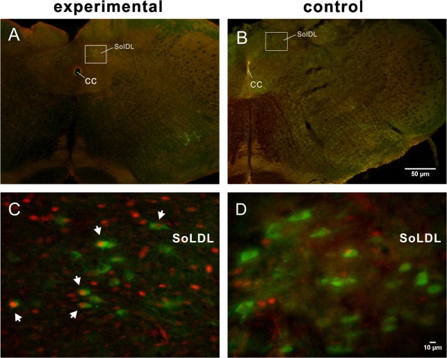
Fig 5. Low-power micrographs of the caudal medulla showing dual-labeled FLI and TH-ir neurons from experimental (A) and control (B) rats at the level of 0.8 mm caudal to the obex.
Dorsal boxed areas in A and B, indicating the SolDL, are enlarged in C and D, respectively, showing double-labeled neurons (white arrows) in the experimental rat but no double labeling in the control rat. Scale bar = 50 μm (applies to A, B), 10 μm (applies to C, D).

