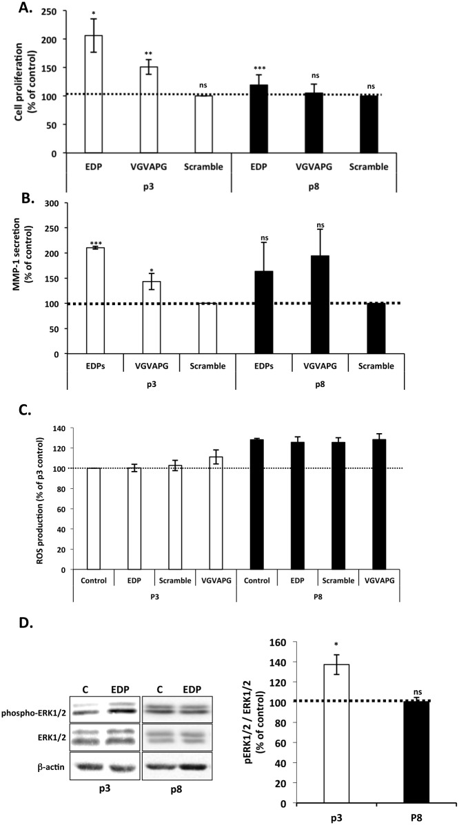
Fig 2. Impact of in vitro aging on EDP biological effects.
Young and aged fibroblasts were represented by passage 3 (white) and 8 (black bars), respectively. Dotted lines (100%) represent the values of the unstimulated controls. (A) Impact of in vitro aging on EDP-mediated fibroblast proliferation. Cells were stimulated or not with 50 μg/mL of EDP or 200 μg/mL of scramble or VGVAPG peptides during 48h. Proliferation was analyzed by counting using Trypan Blue exclusion. (B) Impact of in vitro aging on EDP-induced MMP-1 secretion on fibroblasts. MMP-1 secretion was evaluated by ELISA. Cells were stimulated with 50 μg/mL of EDP or 200 μg/mL of scramble or VGVAPG peptides during 24h. (C) Impact of in vitro aging on EDP-induced ROS production. Cells were stimulated for 30 minutes with 50 μg/mL of EDP or 200 μg/mL of scramble or VGVAPG peptides. ROS were detected by the Image-iT LIVE Green Reactive Oxygen Species Detection Kit as described. (D) Impact of in vitro aging on EDP-induced ERK1/2 activation. ERK1/2 phosphorylation was analyzed by Western blotting after 30 min of stimulation with 50 μg/mL of EDP. Cells extracts were analyzed using anti-phospho-ERK1/2 (T202/Y204) and anti-ERK1/2 antibodies. The blots are presented in the left panel. The corresponding densitometric analysis is provided on the right panel. *, p<0.05, ** p<0.02, *** p<0.01.

