
Fig. 1.
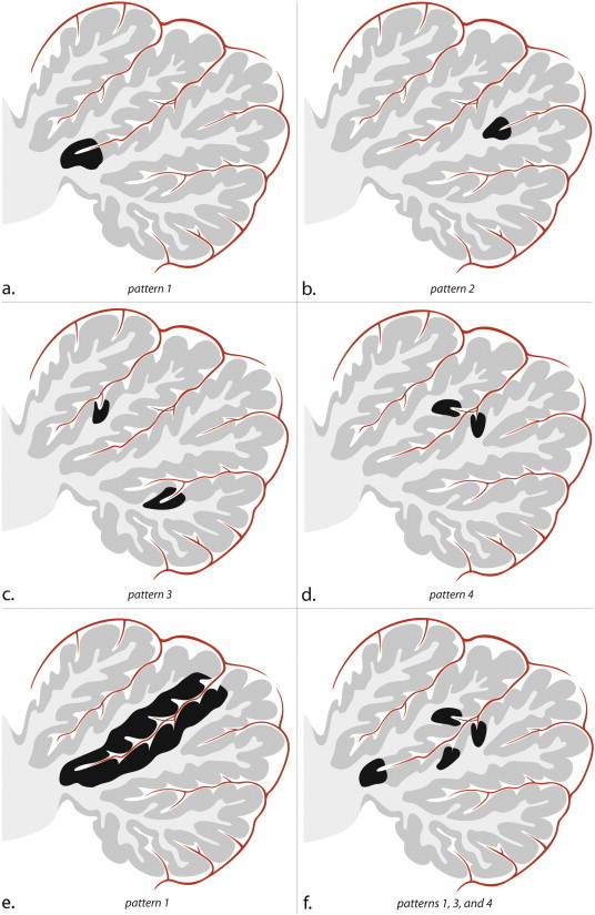
Schematic sagittal drawings illustrate a single cerebellar lobe, its arterial supply, and the patterns of small infarcts. Within the depicted cerebellar lobe, multiple folia are separated by fissures. The folia, which consist of the cortex and subcortical white matter, converge towards the deep white matter of the cerebellum. In the fissures, an arterial branch is present which gives rise to cortical arteries. (a) Pattern 1 corresponds to infarcts involving the apex of a large fissure, (b) pattern 2 corresponds to infarcts involving the apex of a shallow fissure, (c) two infarcts corresponding to pattern 3, and (d) one infarct involving opposite sides of a fissure, indicative of pattern 4. (e) Infarct involving the entire cortical coating of a deep cerebellar fissure; notice this also is a pattern 1 infarct since it involves the apex of a deep fissure. This infarct likely resulted from the occlusion of the arterial branch in the cerebellar fissure. (f) Combinations of small infarcts commonly occur alongside the same fissure. (a–f) Notice the sparing of both subcortical and deep white matter in each cortical infarct.