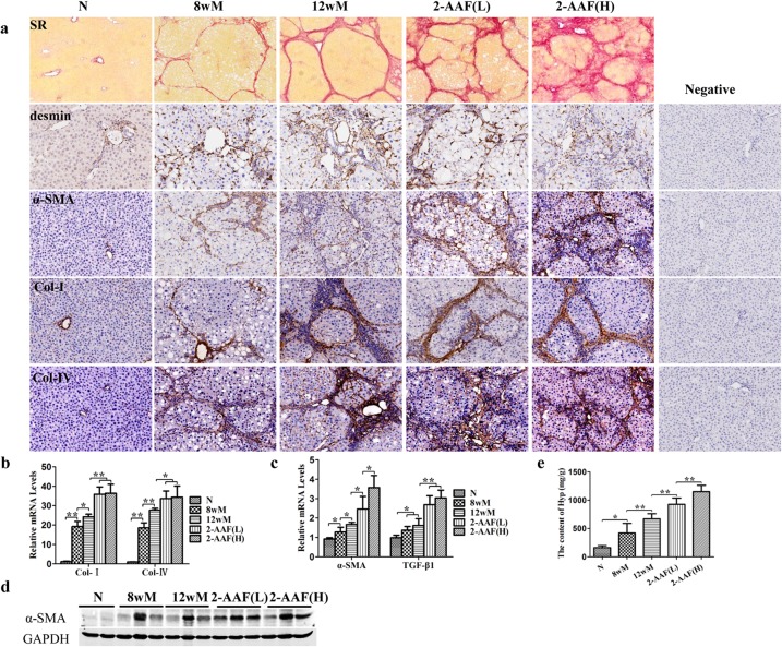
Fig 2. 2-AAF advanced the progress of cirrhosis.
(a) Sirius Red Staining (100×) and immunohistochemistry of desmin, α-SMA, Col I and Col IV (×200). (b) The relative expression levels of Col I and Col IV were measured by qRT-PCR. (c) The relative expression levels of α-SMA and TGF-β1 were measured by qRT-PCR. (d) Western blot of α-SMA. (e) The hydroxyproline content of liver tissue. *p < 0.05, **p < 0.01.

