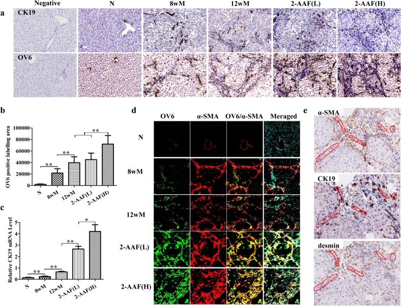
Fig 3. 2-AAF promoted the activation and expansion of HPCs, and promoted the differentiation of HPCs to myofibroblasts.
(a) Immunohistochemistry of CK19 and OV6 (×200). (b) The change in the OV6-positive labeling area. (c) The relative expression level of CK19 was measured by qRT-PCR. (d) Double immune-staining of α-SMA (red), OV6 (green) and DAPI (blue), merged α-SMA, OV6 and DAPI (×200). (e) Serial section immunostaining of α-SMA, CK19 and desmin in the 2-AAF(H) group (×200). *p < 0.05, **p < 0.01.

