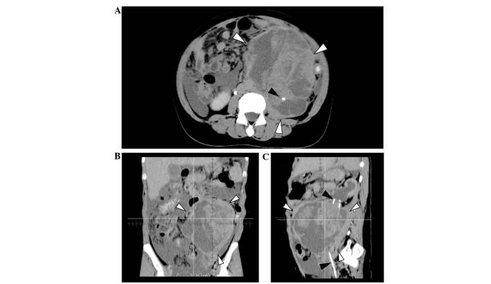
Figure 1.
Computed tomography images prior to treatment. (A) Axial image showing the tumor of 12×16×16 cm located in the left abdomen (white arrowheads). The left ureter is involved in the tumor, and the ureteral catheter is visible in the tumor (black arrowhead). A small amount of ascites was also detected. (B) Coronal image showing the tumor with heterogeneous appearance and including cystic and solid components. (C) Sagittal image: Ureteral catheter penetrating the tumor (black arrowheads) and hydronephrosis were observed.

