
Figure 2.
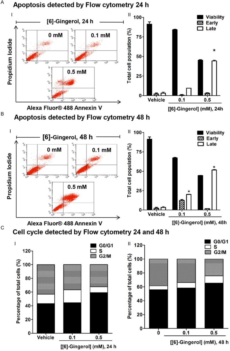
[6]-gingerol promoted apoptosis and cell cycle arrest in HepG2 cells. Cells were treated with 0.1 and 0.5 mM [6]-gingerol for 24 h and 48 h. Cell cycle and apoptosis analysis were determined by flow cytometry. The control was defined as cells treated with a medium or 0.1% DMSO vehicle without [6]-gingerol. AI. Cell death and apoptosis following [6]-gingerol treatment for 24 h identified as the events that were single stained for Alexa Fluor488-Annexin V (lower right quadrant or early apoptisis) or double stained for both Alexa Fluor® 488-Annexin V and PI (upper right quadrant or late apoptosis). II. The distribution of viable, early, and late apoptotic cells were evaluated relative to the whole cell populations (set as 100%). BI. Cell death and apoptosis following [6]-gingerol treatment for 48 h. II. The distribution of viable, early, and late apoptotic cells were evaluated relative to the whole cell populations (set as 100%). C. PI staining was used to determine the percentages of cells in each phase of the cell cycle relative to the whole cell populations (set as 100%) and expressed in histogram profile of cell cycle distribution in Go/G1, S, and G2/M phases after 24 h I and 48 h II of [6]-gingerol treatment. Three independent experiments were performed for statistical analysis and expressed as mean ± SD. *denotes statistically significant difference from the control at P<0.05.