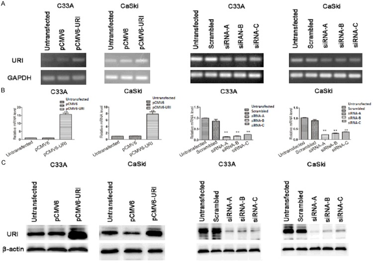
Figure 1.
URI expression in cervical cancer cells. A. The expression level of URI mRNA in transfected cervical cells was examined by RT-PCR. GAPDH served as a loading control. B. Relative mRNA transcripts of URI were examined using qRT-PCR. C. Western blot analysis to confirm the siRNA mediated knockdown of URI and the pCMV6-URI mediated overexpression of URI in C33A and CaSki cells. β-actin was used as internal control. left two panels of A-C: URI over-expression; right two panels of A-C: URI siRNA transfection. Data was expressed as mean ± SEM of three independent experiments, **p<0.01.

