
Figure 2.
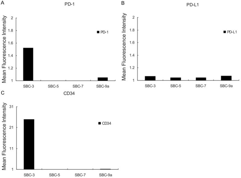
Summary of flow cytometric analysis of SCLC cell lines. A. Anti-human PD-1: PD-1 molecule was positively detected in SBC-3 and SBC-9a cells. The expression levels of PD-1 molecules on SBC-5 and SBC-7 cells were relatively low compared with SBC-3 and SBC-9a cells. Thus, PD-1 molecule was not expressed on the cell-surface of these cell lines. B. Anti-human PD-L1: PD-L1 molecule was weakly expressed on the cell-surface of all cell lines examined in this experiment. C. Anti-human CD34: positive control.