Figure 1.

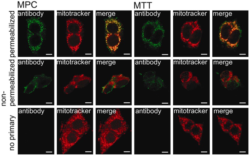
ATP synthase is present on the surface of mouse pheochromocytoma cells. Separate confocal microscopy channels of MPC (left) and MTT (right) cells labeled with ATP5B antibody (green signal) and mitotracker (red signal) and merged channels. The top row shows results for permeabilized cells, the center panel shows non-permeabilized cells, and the bottom panel shows no primary antibody negative controls. Both MPC and MTT showed strong positive staining for ATP5B that greatly, but not entirely, overlapped with the mitochondria (top row). Signal for ATP5B on non-permeabilized cells does not overlap with mitochondrial staining, indicating cell surface location of ATP5B (second row). Application of secondary antibody only did not lead to any detectable signal (bottom row). Scale bar indicates 10 μm.
