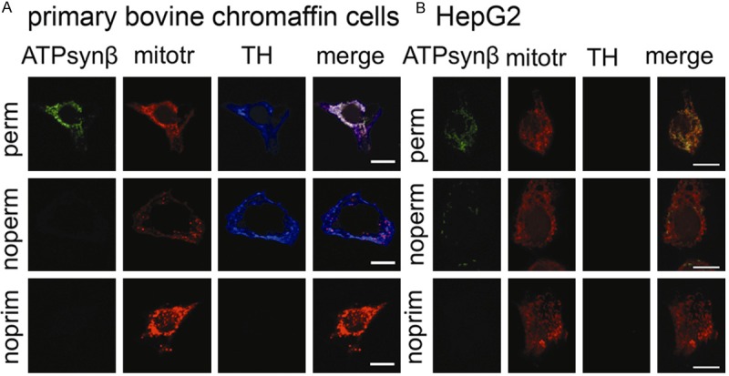
Figure 2.

ATP synthase is absent from the surface of normal bovine chromaffin cells. Confocal images of primary bovine chromaffin cells (A) and HepG2 cells (B) labeled with mouse anti-ATP5B (ATPsynβ) or tyrosine hydroxylase (TH) antibody and mitotracker (mitotr) in permeabilized cells (perm, top row), non-permeabilized cells (noperm, second row), and no primary antibody negative controls (noprim, third row). Merged channels for ATP5B and mitotracker show complete overlap in permeabilized cells (top row), while no signal for ATP5B was evident in non-permeabilized cells (second row) or the negative control (third row) for primary bovine chromaffin cells (A). Positive staining for TH confirms chromaffin cell origin. Non-permeabilized HepG2 cells showed punctate ATP5B signal. As expected, no TH signal was detectable for HepG2 cells. In conclusion cell surface localization of ATP5B was non-detectable in primary chromaffin cells while present on HepG2 cells. Scale bar indicates 10 μm.
