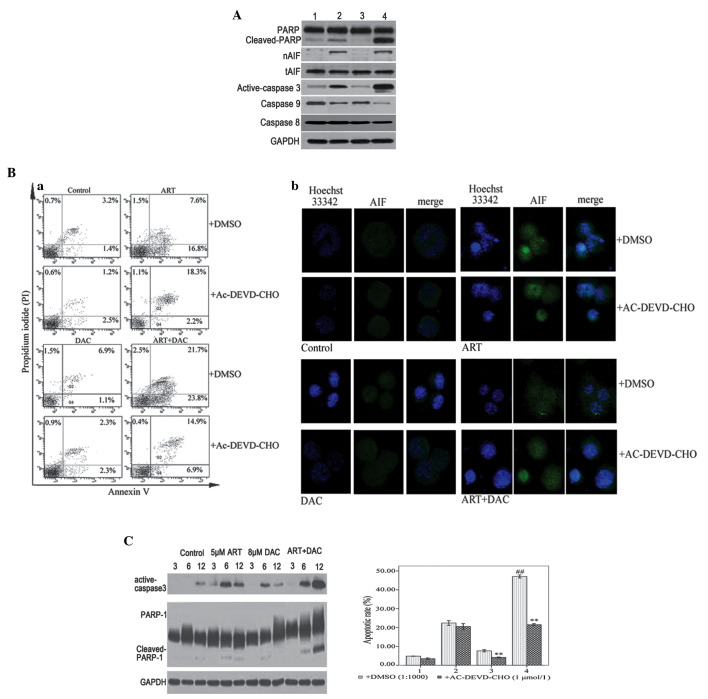
Figure 3.
(A) SKM-1 cells were treated with 8 µmol/l decitabine (DAC) or 0.1% dimethylsulfoxide (DMSO) for 30 min, and then further incubated with 5 µmol/l artesunate (ART) for 12 h. Whole cell lysates or nuclear proteins were analyzed by western blotting for the apoptotic proteins, caspase-9, −8, and-3, poly(ADP-ribose) polymerase-1 (PARP-1) and apoptosis-inducing factor (AIF), with GAPDH as the loading control. Lane 1, control group; lane 2, ART group; lane 3, DAC group; and lane 4, DAC and ART group. (B) (a) SKM-1 cells were treated with 8 µmol/l DAC or 0.1% DMSO for 30 min, and then further incubated with 5 µmol/l ART for 12 h. The Annexin V/propidium iodide staining revealed that compared with single-agent treatment, the combination of ART and DAC induced an increased rate of apoptosis. The caspase-3/7 inhibitor, Ac-DEVD-CHO, abrogated apoptosis induced by DAC, but not ART. (B) (b) SKM-1 cells were treated with 8 µmol/l DAC or 0.1% DMSO for 30 min, and then further incubated with 5 µmol/l ART for 6 h. Laser confocal microscope analysis revealed that ART, but not DAC, induced AIF transfer to the nucleus. (C) SKM-1 cells were treated with DAC and/or ART for 3, 6 or 12 h. Western blot analysis revealed that DAC and ART induced caspase-dependent apoptosis. The presence of active caspase-3 and cleaved PARP, induced by DAC or ART alone, was not observed after 12 h. However, the combined ART-DAC treatment induced a notable activation of caspase-3 and cleaved PARP, which was observed even after 12 h. **P<0.005 compared with the control group treated with DMSO; and ##P<0.005 compared with the other groups.

