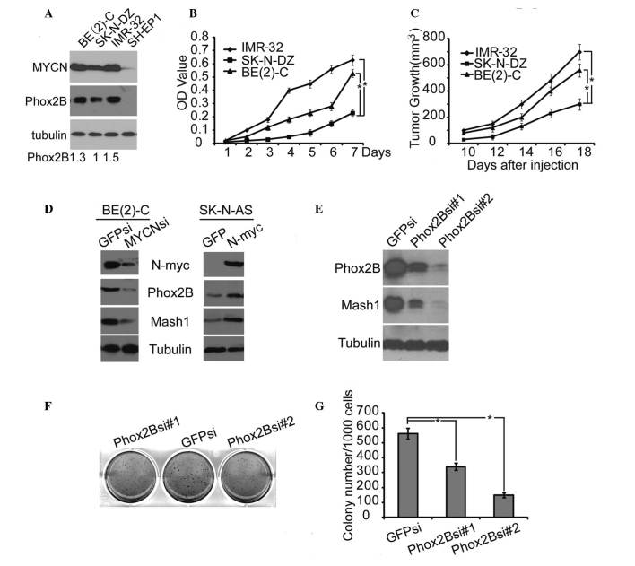
Figure 4.
High level of Phox2B expression promoted human neuroblastoma cell proliferation and xenograft tumor growth. (A) Western blot analysis of MYCN and Phox2B expression in 4 neuroblastoma cell lines. α-Tubulin levels are presented as the loading control; the quantification of the expression level of Phox2B is normalized to that of the tubulin control. (B) Cell proliferation assay of 3 neuroblastoma cell lines, IMR-32, SK-N-DZ, and BE(2)-C, was performed using CCK-8. (C) Tumor growth in NOD/SCID mice injected with the 3 neuroblastoma cell lines, IMR-32, SK-N-DZ or BE(2)-C. (D) Western blot analysis of MYCN, Phox2B and Mash1 expression in BE(2)-C cells with MYCN knockdown by shRNA, or in SK-N-AS cells with MYCN overexpression. (E) Western blot analysis of Phox2B and Mash1 expression in BE(2)-C cells with Phox2B knockdown by shRNA. (F) Soft agar colony formation assay of BE(2)-C cells with Phox2B knockdown by shRNA. (G) Colonies ≥0.5 mm or comprising ≥50 cells were recorded. The data in B, C and G are presented as the mean ± standard deviation, obtained from 3 independent experiments. Statistical analysis was performed using two-tailed Student's t-test, *P≤0.05. Phox2B, paired-like homeobox 2b; MYCN, v-myc avian myelocytomatosis viral oncogene neuroblastoma derived homolog; CCK-8, cell counting kit 8; Mash1, mammalian achaete-scute homolog 1; shRNA, short hairpin RNA.

