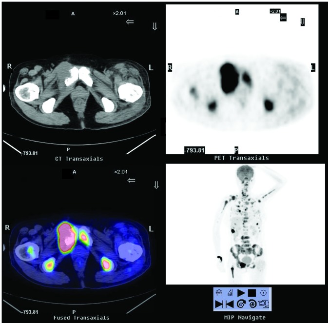
Figure 3.
18F-FDG PET/CT scintigraphy from January 2013, showing more severe bone destruction as well as a notably increased 18F-FDG uptake in most of the previous bone lesions and within multiple pulmonary nodules and lymph nodes. This pointed to the presence of metastatic tumors. 18F-FDG PET/CT, 18F-fluorodeoxyglucose positron emission tomography/computed tomography; A, anterior; P, posterior; R, right; L, left.

