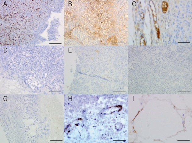
Figure 4.

Immunohistochemistry in murine flank xenografts A&D: Staining for Ki-67 proliferation marker in a U87 murine flank xenograft partially resected and treated with blank poly(lactic-co-glycolic acid)/poly(ethylene glycol) (PLGA/PEG) (A) or PLGA/PEG loaded with 160mg/kg etoposide (D), showing reduction in tumour cell proliferation in response to active etoposide release
B&E: Staining for vascular endothelial growth factor (VEGF) in a U87 murine flank xenograft partially resected and treated with blank PLGA/PEG (B) or PLGA/PEG loaded with 160mg/kg etoposide (E), showing reduction in VEGF positivity
C&F: Staining for the angiogenic vessel marker endoglin, showing new vessel formation in a U87 murine flank xenograft treated with blank PLGA/PEG (C) but not for PLGA/PEG loaded with 160mg/kg etoposide (F)
G: Low power view of an excised U87 murine flank xenograft treated with PLGA/PEG loaded with 160mg/kg etoposide showing the inner zone of necrosis, more viable tumour centrally and the outer capsule of normal mouse tissue
H: Immunostaining for endoglin in a U87 murine flank xenograft treated with PLGA/PEG loaded with 160mg/kg etoposide showing damaged blood vessels in highly necrotic tissue
I: Immunostaining for VEGF with tumour cells growing around blank PLGA/PEG particles showing the non-toxic nature of the carrier biomaterial
