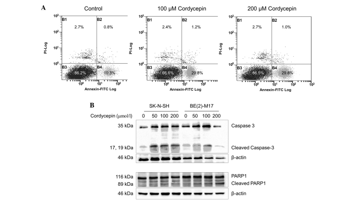
Figure 3.
Cordycepin induces apoptotic cell death in neuroblastoma cells. (A) The SK-N-SH cells were treated with 0, 100 and 200 µmol/l cordycepin for 48 h prior to staining with Annexin V-PI and assessment using fluorescence-activated cell sorting analysis. (B) Western blot analysis was performed to determine the level of apoptotic markers in SK-N-SH cells treated with cordycepin. β-actin was used as a loading control. The bands between the full length 116-kDa and cleaved 89-kDa PARP1 bands are non-specific. PI, propidium iodide; FITC, fluorescein isothiocynate; PARP1, poly[ADP-ribose] polymerase 1.

