Fig. 4.
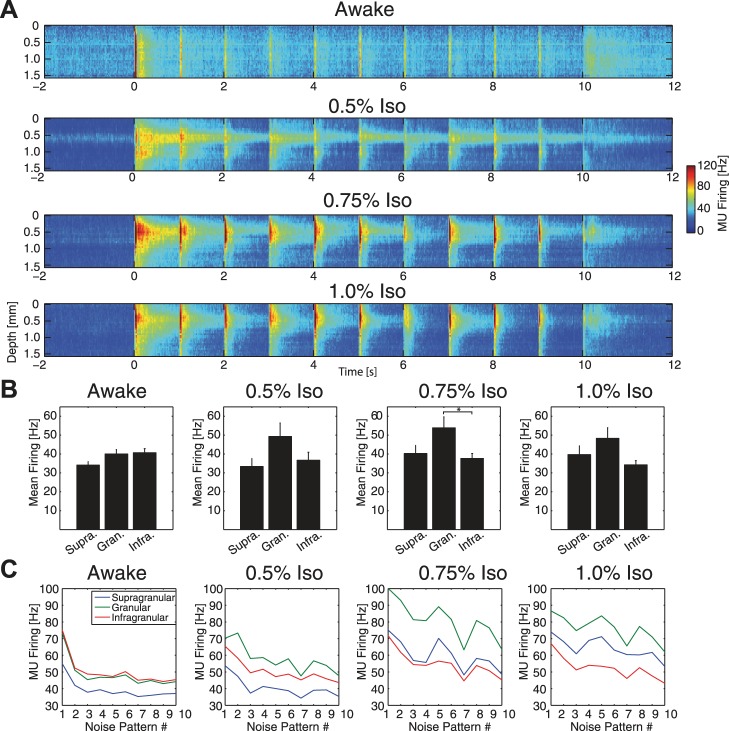
Disruption of the laminar distribution and adaptation of visually-evoked MUA responses. A: group-averaged MU firing rate across cortical layers. Compared with awake animals (top), increasing concentrations of anesthetic (bottom; 0.5%, 0.75%, 1.0% iso, all with xylazine) altered the laminar distribution of MU firing, notably increasing relative strength of the response in putative layer IV (electrode depth 0.3–0.6 mm). B: group-averaged mean firing rate across 10 s of visual stimulation for supragranular, granular, and infragranular layers. Awake animals exhibited slightly higher MU firing rate in granular and infragranular layers compared with supragranular layers. 0.5%, 0.75%, and 1.0% iso all with xylazine increased firing rate in granular layers at the trend level relative to supragranular and infragranular layers. Error bars indicate 1 SE. *Significantly different at P < 0.05. C: response to each of the 10 transitions between subsequent noise patterns during the visual stimulus, calculated as the mean MU firing rate for 200 ms after each screen change, for supragranular (blue), granular (green), and infragranular (red) layers. Awake animals exhibited pronounced spike rate adaptation for later noise patterns, while anesthetics slowed this adaptation of MUA.

