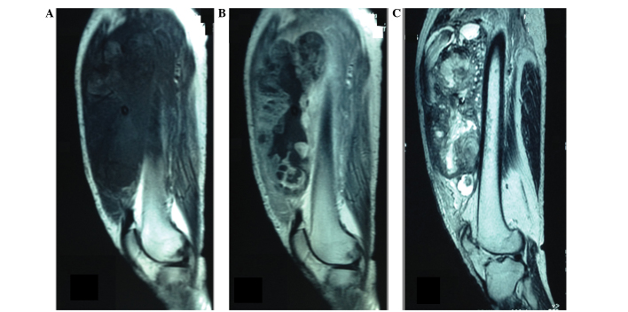
Figure 1.
(A) Sagittal T1-weighted MRI showing a large, irregularly lobulated mass, slightly hypointense to skeletal muscle. (B) Sagittal contrast-enhanced T1-weighted MRI revealing heterogeneous contrast enhancement of the tumor with massive central necrosis. (C) Sagittal T2-weighted MRI demonstrating multiple intratumoral cystic areas. MRI, magnetic resonance imaging.

