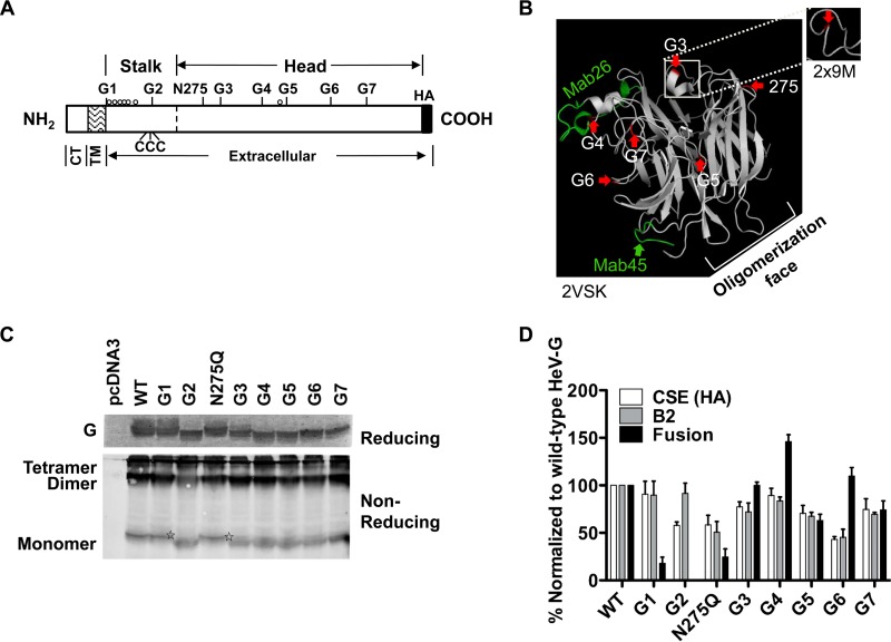
FIG 1.
Individual characterizations of all eight predicted N-glycosylation sites in HeV G. (A) Schematic representation of HeV G, including the positions of the eight potential glycosylation sites. The cytoplasmic tail (CT), transmembrane (TM), and extracellular domains of HeV G are indicated. Three cysteine residues in the stalk domain that are important for G oligomerization are depicted (CCC) (13). O-glycosylation sites published for HeV G are illustrated by open circles (24). (B) N-glycan structural positions in the head of HeV G. Side view of the cartoon representation of the monomeric subunit structure of the HeV G globular head domain, taken from HeV G in complex with ephrin B2 (PDB ID 2VSK) (38). Interestingly, the G3 region from the dimeric crystal structure of HeV G (PDB ID 2X9M) (39) shows this region to form a loop (outer box) instead of an alpha helix (inner box). Structures were attained using PYMOL (www.pymol.org). The structure displays the positions of the predicted HeV G head N-glycosylation sites (G3 to G7 and position 275, highlighted in red and marked by red arrows). The likely oligomerization face of HeV G (39) is indicated. The MAb26 and MAb45 binding regions (11) are highlighted in green. (C) 293T cells were transfected with WT or mutant HeV G expression constructs and lysed 20 to 24 h posttransfection. Lysates were analyzed for HeV G expression by reducing (top gel) or nonreducing (bottom gel) PAGE and subsequent immunoblotting against the C-terminal HA tag in HeV G using rabbit anti-HA antibody. The reducing gel resolved the monomeric form of HeV G, whereas the nonreducing gel also resolved the dimeric and tetrameric forms of HeV G, as indicated. Stars denote the monomeric G of the unoccupied N-glycosylation sites of the G1 and N275Q mutants, running with the same mobility as WT HeV G. (D) 293T cell surface expression (CSE) and ephrin B2 (B2) receptor binding of HeV G N-glycan mutants was determined by flow cytometry using mouse anti-HA antibody or soluble mouse ephrin B2/Fc chimeric protein, respectively. The fusion promotion abilities of the HeV G N-glycan mutants were assessed in 293T cells by cotransfection with WT HeV F. Syncytial nuclei were counted as outlined in Materials and Methods. The CSE, ephrin B2 binding, and fusion levels of the HeV G N-glycan mutants were normalized to those of WT HeV G. Data shown are the average results ± standard errors of the means (SEM) from at least three independent experiments.

