FIG 1.
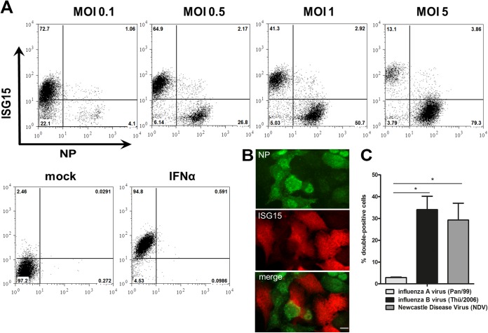
ISG15 is predominantly induced in noninfected cells upon IAV infection. (A) A549 cells were infected with the seasonal Pan/99 strain at the indicated MOI. At 24 h postinfection (hpi) cells were stained for ISG15 (y axis) and NP (x axis) and analyzed via FACS. Numbers indicate the percentages of cells in each gate. One representative dot plot is shown for each MOI (n > 3). Control staining is shown for cells after mock infection or treatment of cells with 500 U/ml IFN-α. (B) A549 cells were infected with Pan/99 at an MOI of 1. At 16 hpi, cells were stained for ISG15 (red channel) and NP (green channel) and analyzed via indirect immunofluorescence assay. One representative image is shown in each panel (n = 2). Bar, 10 μm. (C) Percentages of ISG15- and NP-double-positive cells at 24 hpi with Pan/99 (influenza A virus; light gray), Thü/2006 (influenza B virus; black), or NDV (Newcastle disease virus; dark gray) when more than 80% of the A549 cells in the respective experiment were positive for viral antigen. Data are means + standard errors of the means (SEM) (n = 4), *, P < 0.05 (Mann-Whitney U test).

