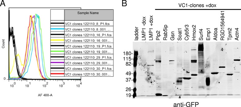
FIG 2.
VC1 clone BiFC and fusion protein expression. (A) VC1 clones were tested for BiFC induction by flow cytometry. Clones were plated in duplicate and tested for BiFC following overnight induction with Dox at 500 ng/ml. The YFP fluorescence of uninduced cells (black histograms) was compared to that of induced cells (colored histograms). AF, Alexa Fluor. (B) Induced clones were examined for CYFP-fusion proteins by Western blotting with a GFP-specific monoclonal antibody that specifically recognizes the CYFP domain. Uninduced and induced control cells with BiFC (LMP1 without Dox and LMP1 with Dox, respectively) and the ladder are indicated. Control cells tested for BiFC inducibly express LMP1-NYFP and LMP1-CYFP. Numbers on the left are molecular masses (in kilodaltons).

