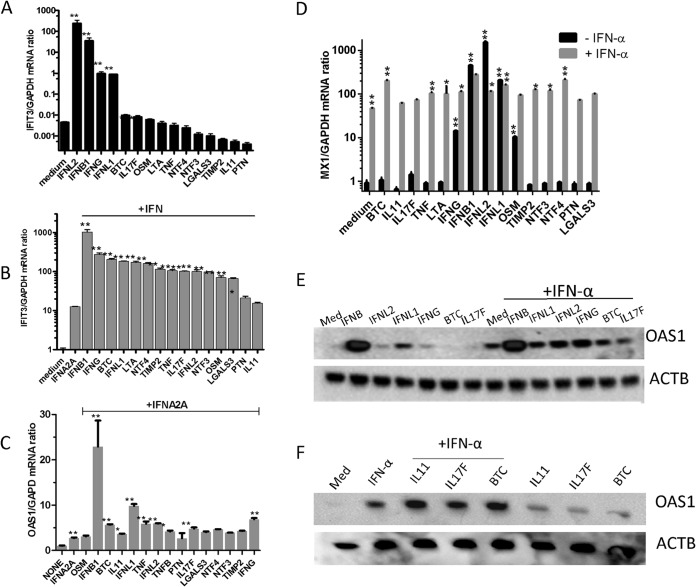
FIG 4.
Induction of ISG expression by the cytokine cluster. (A) Huh-7 cells were pretreated with selected cytokines (∼1 nM) or a combination for 6 h. Quantitative RT-PCR using TaqMan primers specific for human IFIT3 and control GAPDH was performed. (B and C) Huh-7 cells were treated with IFN-α (10 IU/ml) with or without selected cytokines (∼1 nM) for 6 h. Quantitative RT-PCR using TaqMan primers specific for human IFIT3 (B) or for human OAS1 (C) was performed, while GAPDH primers were used for GAPDH housekeeping control. (D) Huh-7 cells were treated with cytokines individually or in combination with IFN-α (10 IU/ml) for 6 h. Quantitative RT-PCR using TaqMan primers specific for human MX1 and GAPDH mRNA levels was performed. The values shown represent the mean ISG/GADPH mRNA ratio plus SEM from two independent experiments. **, P < 0.01. (E and F) Huh-7 cells were treated with IFN-α (10 IU/ml) with or without selected cytokines (∼1 nM). The Huh-7 cells were treated with the indicated cytokine combinations for 20 h, and then lysates (50 μg) were obtained and resolved by SDS-PAGE. Western blots were performed using an OAS1 antibody. The blots were reprobed with an anti-β-actin antibody for loading control. The images are from a representative experiment out of two. *, P < 0.01; **, P < 0.001.

