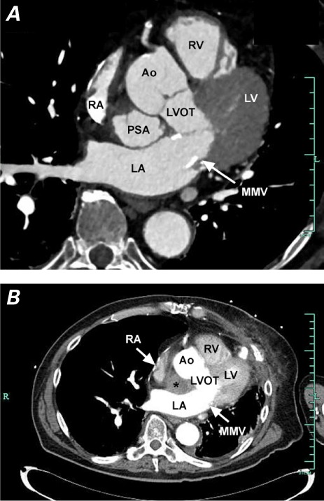
Fig. 2.

Computed tomographic angiograms show the pseudoaneurysm A) untreated, in the mitral-aortic intervalvular fibrosa; and B) with no contrast entry, indicating successful occlusion.
* = occluded pseudoaneurysm; Ao = aorta; LA = left atrium; LV = left ventricle; LVOT = left ventricular outflow tract; MMV = mechanical mitral valve; PSA = pseudoaneurysm; RA = right atrium; RV = right ventricle
