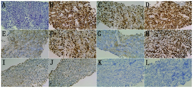
Figure 4.
Diffuse infiltration of large B-cells. (A) Hematoxylin and eosin stained cells. Immunohistochemical analysis revealed that the samples were positive for (B) CD20, (C) CD79a, (D) BCL-6, (E) CD10, (F) MUM-1, partially positive for (G) EMA and (H) Ki-67 index 70%; and negative for (I) CD5, (J) CD3, (K) BCL-2 and (L) AE1/AE3. (hematoxylin and eosin staining; magnification, x200). MUM-1, multiple myeloma oncogene 1; EMA, epithelial membrane antigen; Bcl, B-cell lymphoma; AE, anion exchanger.

