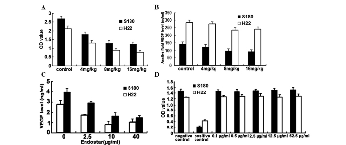
Figure 2.
(A) Results of the peritoneal permeability assay in the S180 and H22 mouse models. Endostar (4, 8 and 16 mg/kg) significantly decreased the permeability of the peritoneum (all P<0.05 vs. control group). (B) Results of the ELISA for VEGF levels in ascites. The cell-free ascites fluid was collected from the peritoneal cavity of the experimental mice. The 8 and 16 mg/kg Endostar-treated groups exhibited marked differences in the level of VEGF (P<0.05 vs. control group). (C) Results of the ELISA for in vitro VEGF secretion in S180 and H22 cells. In total, 1×10⁴ cells/well were treated with various concentrations of Endostar (0, 2.5, 10 and 40 µg/ml) for 48 h. The media from the wells was then individually collected and analyzed. Endostar reduced VEGF secretion (all P<0.05 vs. control group). (D) Results of the in vitro MTT proliferation assay for S180 and H22 cells. The S180 and H22 cells were treated with Endostar at various doses (0.1, 0.5, 2.5, 12.5 and 62.5 µg/ml). Endostar did not reduce the proliferation of S180 and H22 cells (all P>0.05 vs. control group). The data are expressed as the mean ± standard deviation. VEGF, vascular endothelial growth factor; OD, optical density.

