
Fig. 8.
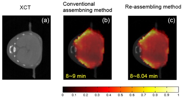
The reconstruction results from the dynamic in vivo experiment, obtained by using the conventional and the re-assembled measurement data. (a) The x-ray computed tomography image of liver region. (b) and (c) The corresponding fluorescence tomographic images that are imaged about 8 min after ICG injection. (b) The reconstruction results obtained by using the conventional assembled measurement data (120-frame). (c) The reconstruction result obtained by using the re-assembled measurement data (2880-frame). The conventional assembled or re-assembled measurement sequences are reconstructed by the KL-based method. The volume considered for reconstruction is a 3-D region and sampled to voxels. The 5,466 voxels inside the imaged object and 19,980 source-detector pairs are used in the reconstruction process of KL domain. The same imaging model is used in the two assembling modes. To improve the reconstruction quality in in vivo experiment, the reconstruction is performed incorporating a heterogeneous forward model. All images are displayed at the same range.