Figure 1.

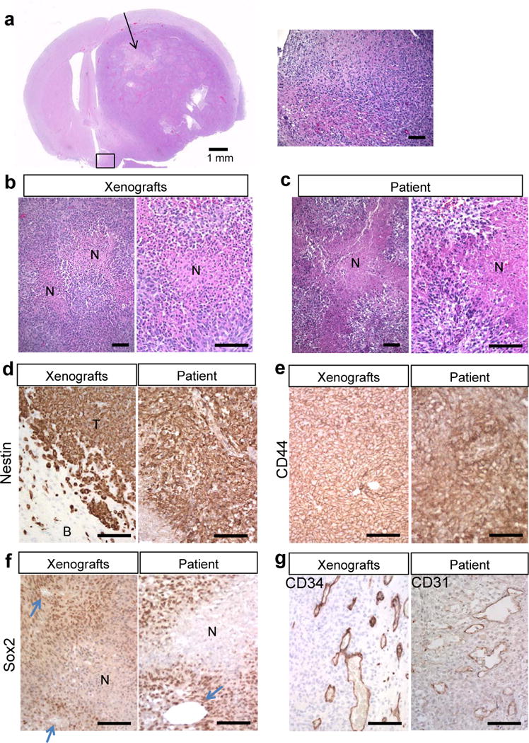
Orthotopic MGG123 xenografts recapitulate the histopathological characteristics of the patient glioblastoma (GBM). (a) Low magnification of an H&E-stained section of a mouse brain bearing a MGG123-derived intracerebral xenograft (left panel). Arrow indicates a large area of necrosis. Higher-magnification of the boxed area on the left panel showing ill-demarcated tumor brain interfaces and tumor invasiveness (right panel). (b, c) H&E staining of orthotopic MGG123 xenografts (b) and the patient tumor (c) showing necrotic foci with palisades. N, necrosis. (d–g) Immunohistochemical characterization of the MGG123 model and the original GBM tissue. Positivity is indicated by brown. Staining for human nestin (d) shows positivity in tumor cells in both xenografts [T] and the patient’s specimen. Surrounding mouse cells in the brain [B] are negative. CD44 is homogeneously positive in both tumor tissues (e). Sox2 positivity is prominent at perinecrotic and perivascular areas in the xenografts and patient. Arrows point to blood vessels (f). CD34 staining reveals tortuous and dilated vasculature at the tumor periphery in the xenografts (g, left panel). CD31 staining of the patient section reveals similar vasculature (g, right panel). Scale bars: a, left panel, 1 mm; all other panels, 100 μm.
