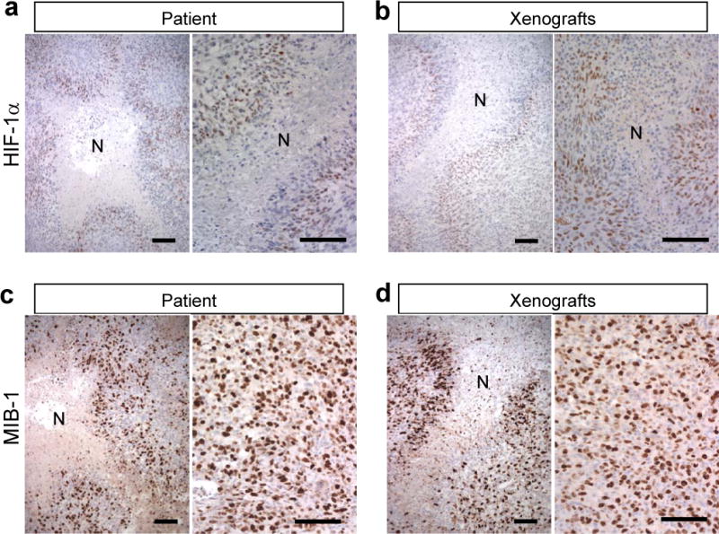
Figure 2.

Perinecrotic neoplastic cells express hypoxia inducible factor 1α (HIF-1α) and are proliferative in the MGG123 model and the patient glioblastoma (GBM). (a, b) HIF-1α immunostaining shows HIF-1α expression (brown) in cells surrounding necrotic foci [N] in both the xenograft and patient tumor. (c, d) MIB-1 is highly expressed in perinecrotic areas as well as other viable tumor areas in both xenografts and patient tumor (labeling index 48% and 44%, respectively). Scale bars, 100 μm.
