
Figure 3.
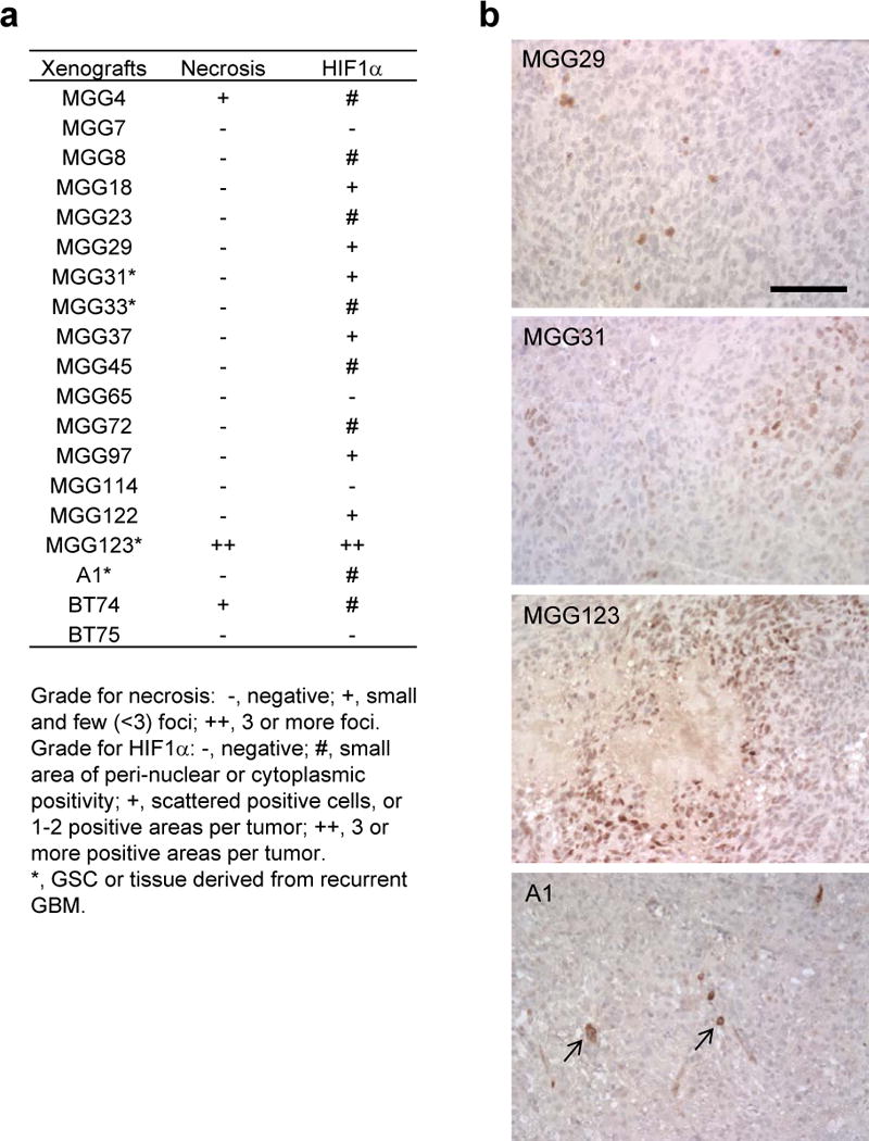
Hypoxia inducible factor 1α (HIF-1α) and necrosis status in a cohort of patient-derived orthotopic glioblastoma (GBM) xenografts. (a) Summary of the status of necrosis and HIF-1α immunohistochemistry (IHC) in 19 patient-derived orthotopic GBM xenografts. (b) Examples of HIF-1α IHC on orthotopic GBM xenografts. Some tumors contain a small number of cells that exhibit HIF-1α staining in the perinuclear areas or cytoplasm (arrows). Scale bars, 100 μm.