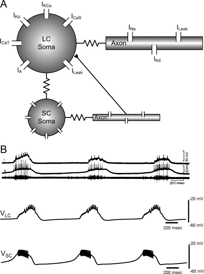
Figure 1.
A, Schematic of the coupled large- and small-cell model. Resistances between soma and axon compartments represent cytoplasmic (axial) resistance, and the resistance between the large- and small-cell somata represents the electrotonic coupling between the two types of cells. EPSPs in the large cell resulting from small-cell action potentials have been implemented with an excitatory chemical synapse linking the small-cell axon to the large-cell soma (filled triangle). B, Comparison of biological and model CG bursting for the network model. Top, Spontaneous bursting activity in P. sanguinolentus as reported by Tazaki and Cooke (1979a). Top trace, Intracellular recording of an anterior large cell. Middle trace, Small-cell recording. Bottom trace, Extracellular recording from main trunk of CG showing action potentials from large cells (large spikes) and small cells (shorter spikes). Bottom, Spontaneous bursts generated in the synaptically coupled large- and small-cell models. Top trace, Large-cell soma voltage. Bottom trace, Small-cell soma voltage.

