



Abstract
Introduction
There are few reports of injury to the soft palate and retropharynx sustained during intubation with the GlideScope® video laryngoscope. Most reports are of isolated injury to the soft palate.
Case History
We describe a patient in whom the retropharynx was injured but the extent of the injury was not observed initially. The patient did not suffer severe sequelae from this injury. However, this injury can cause serious sequelae if it is not recognised (eg development of a retropharyngeal abscess).
Conclusions
We recommend that any patient who sustains injury to the soft palate during intubation (particularly if the endotracheal tube passes through the soft palate) should be reviewed an otolaryngologist before removal of the endotracheal tube.
Keywords: GlideScope® video laryngoscope, Endotracheal tube, Intubation – Palate
The GlideScope® video laryngoscope (GVL) is used to aid the intubation of difficult airways. The system consists of a laryngoscope with a tip containing a video chip, which allows visualisation of the laryngeal inlet and aids accurate placement of an endotracheal tube (ETT). During insertion of the ETT into the mouth, injury may occur within the oral cavity or pharynx if the operator is preoccupied with viewing the video monitor.
Studies have suggested a prevalence of complications of 1% for minor injuries and 0.3% for more serious injuries during insertion of the ETT into the mouth. 1 Several case reports have focused on perforation of the soft palate or one of the pharyngeal arches, and one report has focused on hypopharyngeal injury.
Here, we describe a 44-year-old male who presented for laparoscopic abdominal surgery and sustained injuries to the soft palate and retropharynx. A literature search (Medline) revealed 14 cases of injury to the upper aerodigestive tract secondary to use of the GVL. Of those, 13 were injury to the soft palate only, with one injury to the hypopharynx. No reports of injury to the retropharynx were noted.
Case History
The patient was admitted for laparoscopic cholecystectomy. He was classified Mallampati grade 2 for intubation. The GVL was used for intubation of the patient with a size-8.5 ETT. The rigid stylet of the GVL was in situ. Distal end of the ETT was seen to enter the larynx on the video screen. An oropharyngeal airway was used before intubation.
Upon withdrawal of the GVL blade, the ETT was seen to have passed through the right lateral soft palate. We were the on-call otolaryngology service and were contacted for review. The ETT was seen to pass into the soft palate superior to the right palatine tonsil. Minimal bleeding was noted, so it was decided to continue with the original procedure and undertake repair of the soft palate once the general surgical procedure was complete.
Once the general surgical procedure was completed, the original ETT was withdrawn partially and a new ETT passed using a McCoy blade, thereby securing the airway for removal of the old ETT and repair of the injury.
Examination with a Boyle–Davis mouth gag (Fig 1) revealed injury to the soft palate superior to the right tonsil. Tip of the ETT could not be seen exiting the palate. However, digital palpation of the pharynx revealed that the ETT had remained below the mucosa and traversed the retropharyngeal space. A rigid operating laryngoscope was used for further evaluation. Tip of the ETT was seen emerging from the retropharynx ≈1cm superior and lateral to the left arytenoid cartilage (Fig 2).
Figure 1.

Initial view. The endotracheal tube is seen entering the soft palate (*) superior to the right tonsil (+). The tongue (*) is pushed to the left.
Figure 2.

View of the posterior larynx. The cuff of the initial endotracheal tube (*) can be seen emerging from the mucosa superior and lateral to the left arytenoid (*).
Bleeding was minimal so it was felt that the ETT could be removed safely. After removal, the defect remained (Fig 3). The palatal injury was sutured, whereas the lower (pharyngeal) injury was left unsutured to allow drainage of bleeding or collections.
Figure 3.
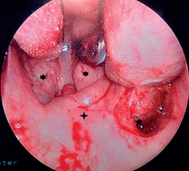
Defect after removal of the endotracheal tube (*). This defect was sutured. Tonsils (*) and soft palate (+) can be seen.
The patient remained intubated overnight in the intensive care unit. He was administered dexamethasone and antibiotics (i.v.). After extubation, he was kept nil-by-mouth for a further 24 hours before starting progressive oral intake. Examination with a flexible nasendoscope was carried out every day during hospital admission. Before discharge from hospital, examination showed that the palatal injury and exit point of the ETT were nearly healed.
From the original procedure, the patient could have been discharged the next day. However, due to the injury, he remained an inpatient for an extra 4 days. He also received 1 week of treatment with antibiotics that would not have otherwise been required. An incident form was completed after the injury. The incident was discussed with the patient and his family. The patient was reviewed in clinic 6-weeks later, by which time the injury had healed fully.
Discussion
A literature search revealed 13 reports of injury to the soft palate and one of injury to the hypopharynx when using the GVL for intubation. This is the first case report in which the injury resulted in an extended stay in hospital.
This case report demonstrates that more extensive injuries can occur using the GVL. The full extent of trauma became apparent only when visualisation of the distal end of the ETT before removal was attempted. This observation suggests that, with any perforation of the soft palate sustained from ETT insertion, further injuries should be assessed before full removal given that the patient can be ventilated safely for this to occur. Ideally, such assessment should be done by an otolaryngology team in case injuries (and their possible sequelae) require management.
A potential serious complication is formation of a retropharyngeal abscess. Such an infection in the deep neck space carries with it a risk of death (estimated to be ≈1%) the risk of developing mediastinitis (estimated mortality ≈50%). To avoid such sequelae occurring in our patient, initially he was kept nil-by-mouth. Before initiating oral intake, the pharynx was examined to assess the wound. Progressive oral intake (starting with sterile water) was commenced after 24 hours because the wound appeared to be healing. Had this not been the case, the patient would have required insertion of a nasogastric tube. The patient was examined every day by nasendoscopy to assess the pharynx and to exclude surgical emphysema. Signs of developing infection were carefully looked for. Broad-spectrum antibiotics were also started.
Several contributory factors can increase the risk of injury/perforation if introducing or passing an ETT. Several strategies have been suggested to reduce this risk: 1–5 altering the way the ETT and stylet are introduced into the oral cavity; ensuring the ETT is visualised passing directly into the oral cavity before watching the laryngoscope monitor; stopping and reassessing if resistance is felt to passage of the ETT (though, in this case, discussion with the anaesthetic team suggested that undue resistance was not encountered); using a softer malleable stylet within the ETT. In this case, it was postulated that use of the oropharyngeal airway before intubation may have caused an initial laceration that the ETT entered and worsened, which would suggest that careful assessment of the oral cavity after using such an adjunct airway is appropriate.
If an injury occurs, we also recommend ensuring that where the tip of the ETT lies is clear, and to ascertain if injury beyond the soft palate has occurred before removal of the ETT. Such confirmation requires referral and review by an otolaryngologist. Given the potential gravity of complications arising from this form of injury, informing patients of the risk of serious injury if intubation using the GVL is considered would seem prudent.
References
- 1. Cooper RM. Complications associated with the use of the GlideScope video laryngoscope. Can J Anaesth 2007; 54: 54–57. [DOI] [PubMed] [Google Scholar]
- 2. Cross P, Cytryn J, Cheng KK. Perforation of the soft palate using the GlideScope video laryngoscope. Can J Anaesth 2007; 54: 588–589. [DOI] [PubMed] [Google Scholar]
- 3. Leong WL, Lim Y, Sia AT. Palatopharyngeal wall perforation during Glidescope intubation. Anaesth Intens Care 2008; 36: 870–874. [DOI] [PubMed] [Google Scholar]
- 4. Malik AM, Frogel JK. Anterior tonsillar pillar perforation during GlideScope video laryngoscopy. Anesth Analg 2007; 104: 1,610–1,611. [DOI] [PubMed] [Google Scholar]
- 5. Vincent RD Jr, Wimberly MP, Brockwell RC, Magnuson JS. Soft palate perforation during orotracheal intubation facilitated by the GlideScope video laryngoscope. J Clin Anesth 2007; 19: 619–621. [DOI] [PubMed] [Google Scholar]