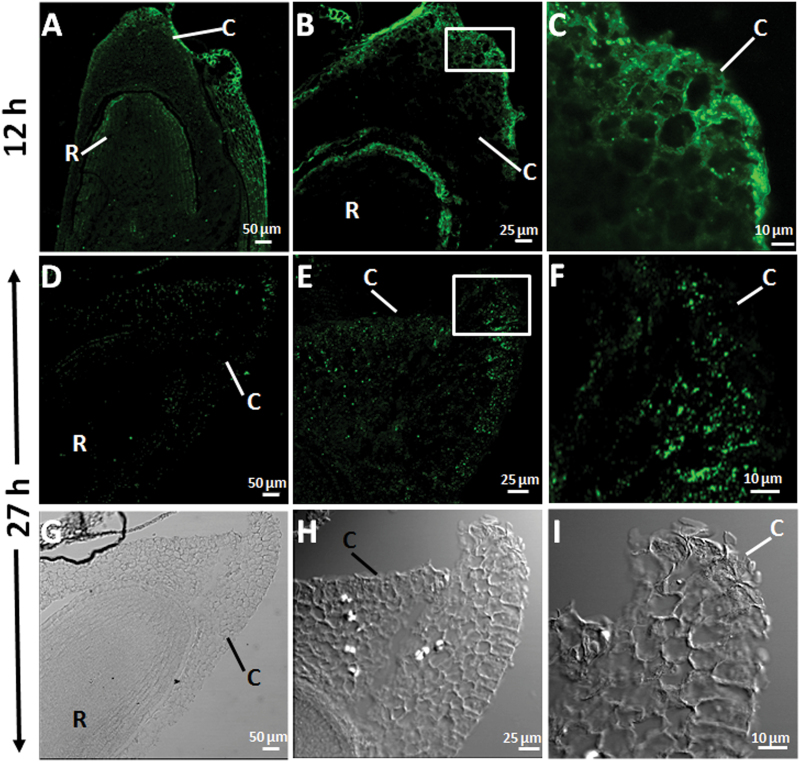
Fig. 2.
Mannan polymer immunolocalization at the root tip and the coleorhiza in longitudinal sections of Brachypodium germinating embryos at (A–C) 12h and at (D–F) 27h. (G–I) DIC images of D, E and F. (C, F, I) Close-up of the coleorhizae. C, coleorhiza; R, root. Scale bars: (A, D, G), 50 μm; (B, E, H), 25 μm; (C, F, I), 10 μm.

