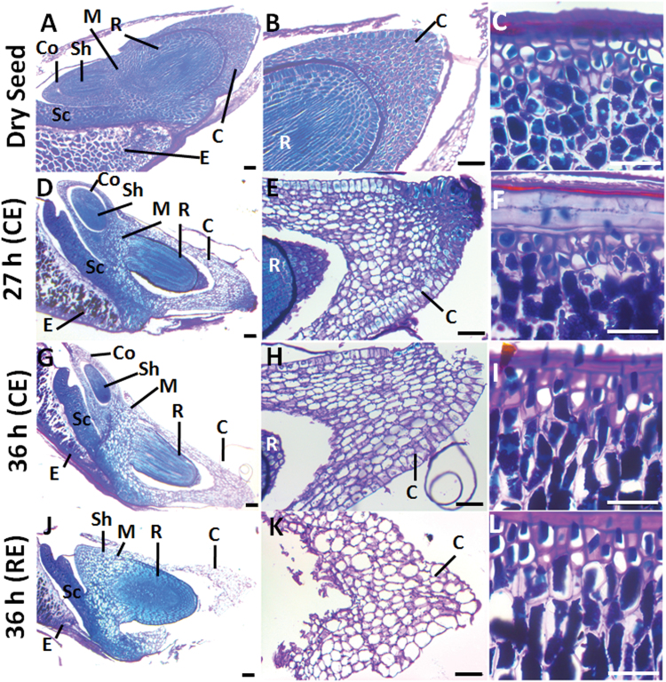
Fig. 7.
Polysaccharide and protein mobilization upon B. distachyon seed germination. Bright field microscopy of longitudinal seed sections stained with PAS-Naphthol Blue Black. (A, D, G, J) Longitudinal sections from dry and water-imbibed seeds at 27h and 36h. (B, E, H, K) Close-up of the coleorhiza in A, D, G, J and (C, F, I, L) close-up of the endosperm in A, D, G, J, respectively. Proteins stain in blue and polysaccharide-rich cell walls in pink. C, coleorhiza; Co, coleoptile; E, endosperm; M, mesocotile; Sc, scutellum; Sh, shoot; R, root. Scale bar: 50 μm.

