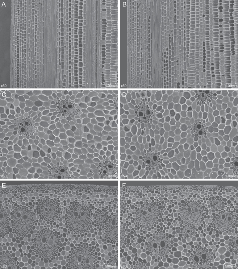
Fig. 3.
Scanning electron microscopy examination of the second internodes from RIL88(qph1) and RIL88(QPH1). The second internodes of near-isogenic lines in mid-elongation stage were subjected to histological analysis. (A, B) Longitudinal view of the parenchyma cells of RIL88(QPH1) and RIL88(QPH1). (C, D) Transverse view of the parenchyma cells of RIL88(qph1) and RIL88(QPH1). (E, F) Transverse view of the epidermal region of RIL88(qph1) and RIL88(QPH1).

